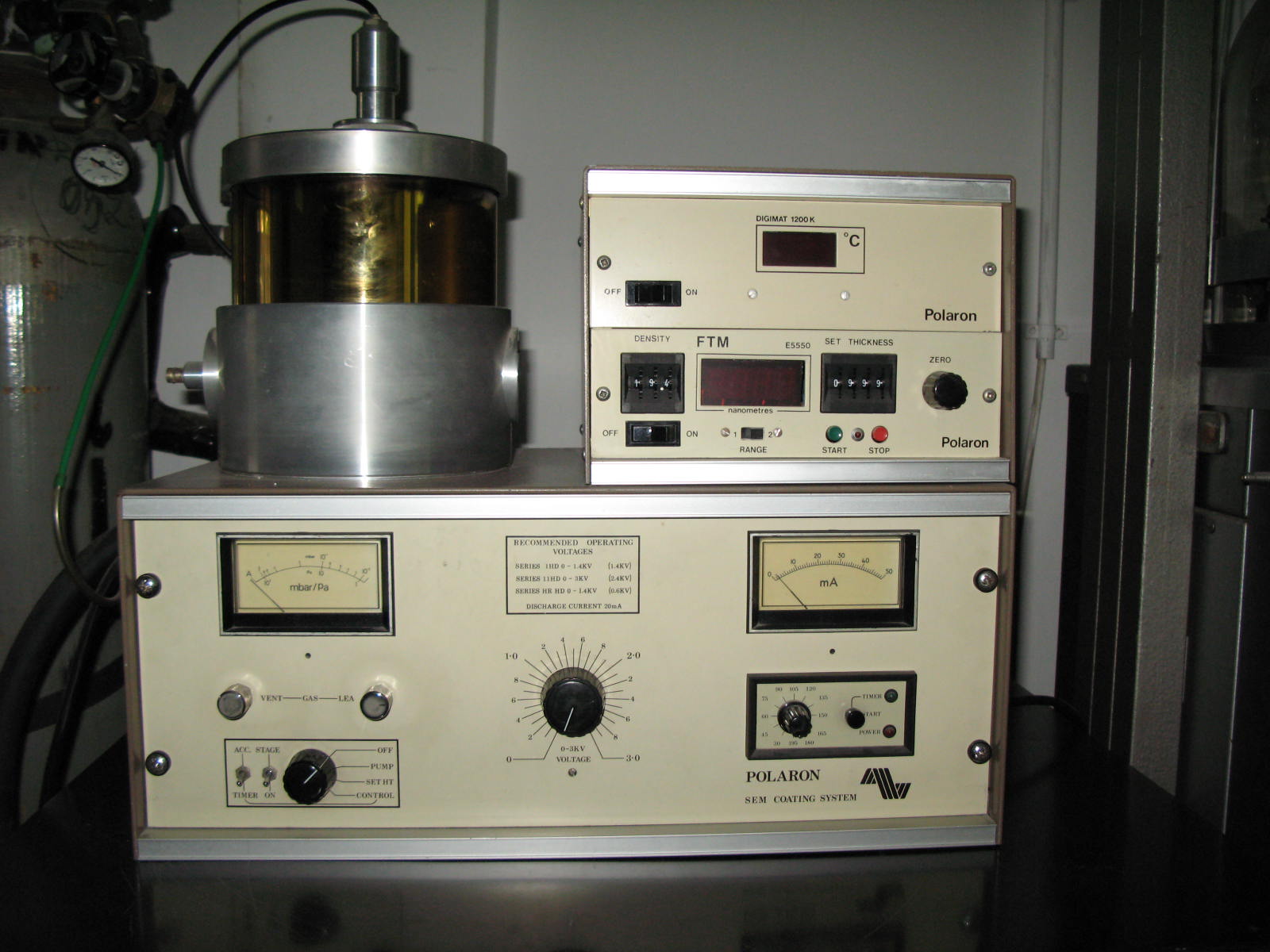
The Interdepartmental Equipment Facility
Gold Sputter Coater

Imaging a sample in the scanning Electron microscope depends on the sample being able to emit 'secondary electrons' in response to the probing electron beam of the SEM. Specimens must also be electrically conductive to facilitate the scanning process. Electrons must have a path to ground to prevent the buildup of static electricity.
Biological specimens, composed largely of carbon compounds, are usually poor conductors and poor emitters of secondary electrons, due to the low atomic number of carbon. Rather than absorbing electrons from the electron source of the microscope and then emitting electrons for detection, carbon compounds tend to collect a charge.
Coating such a specimen with a very thin layer of a heavy metal, such as gold, increases its ability to conduct electricity and emit secondary electrons when in a scanning electron microscope. The gold coating acts as a "stain" for electron microscopy and also increases the apparent contrast so that better images are generated.
The Polaron Sputter coater uses a gold target which is bombarded by fast particles. The target is gradually eroded by the attack and gold atoms are sputtered around. Some of the sputtered atoms condense on the surface of the specimen and form a thin coating.
For effective sputtering to occur the right conditions must exist in the sample chamber. This chamber is flushed with argon at low pressure and has a DC voltage established between the two oppositely set electrodes. When this voltage exceeds the breakdown potential, a self-sustaining glow discharge occurs, characterized by a luminous glow. The gold target, at the cathode is constantly bombarded by ions causing erosion and sputtering. The object to be coated is connected to the anode.