The Interdepartmental Equipment Facility
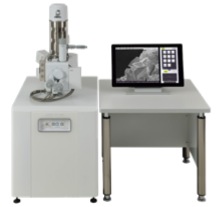
Low vacuum SEM - Scanning Electron Microscope

The Jeol It 100 includes a secondary electron detector and a backscattered detector. It has a high vacuum and low vacuum mode. This instrument is used for millimeter to micron scale samples of dry and semi dry samples.
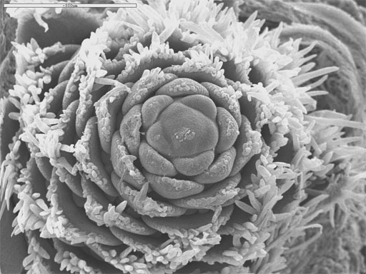
Imaging by SEM. The scanning electron microscope generates an image of the sample surface by probing it with a high-energy beam of electrons. These 'primary' electrons hit the sample and eject secondary electrons. The SEM's primary imaging method is based on this secondary electron emission. These electrons are focused on a scintillation material which generates a flash of light whenever hit by an electron. The light flashes are then detected and amplified by a photomultiplier tube.
The primary electron beam scans the sample point by point, generating secondary electrons wherever it hits. As the intensity of secondary electron emission varies from point to point on the sample, so does the intensity of light emission induced by these electrons . The image obtained is created on a Cathode Ray Tube Screen or an equivalent device. Back-scattered electrons that are reflected from the sample may also be used as signal to form an image.
Resolution and magnification. The wavelength of light rays is the limiting factor in microscopy. A light microscope which uses wavelengths between 400 to 700 nm can distinguish between two adjacent objects separated by ~ 200 nm, and is limited to about 1000-fold magnifications. In contrast, the SEM which uses high energy electrons as its source of 'illumination' with wavelengths of about 0.01 nm is capable of up to 1 million-fold magnifications. Our 5410 SEM is usually used at magnifications between 20 and 35,000 X and can resolve two objects 3.5 nm apart, when operated at 35,000 kV.

Sample Preparation. Air molecules block and scatter the stream of electrons aimed at the sample. This is why electron microscopes require high vacuum for operation. On the other hand, if we want to observe biological samples we have to dry them before putting them in the SEM, to prevent damage by water to the microscope. To overcome this we use the CPD - an instrument which can effectively dry samples with minimal damage to them.
Samples are usually fixed at low temperatures in special fixatives, in order to preserve the morphology and structural integrity of the specimen. The preserved specimen is next sputter-coated with gold, in order to give it the electrical conductivity required for imaging by SEM.
Unique Advantages of the SEM include, 1. high resolution with relatively little sample preparation, 2. large depth of field, which allows work with rough samples, and 3. rapid elemental analysis by (EDS).
