The Interdepartmental Equipment Facility
We've moved to a new Website >>>
Confocal Microscope

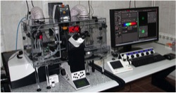
The Leica SP8 Confocal Laser Scanning Microscope includes two PMTs, High resolution photon counting detector (HyD), super galvo stage. It has x10-x63 objectives and cover light spectrum of 400-700 spectral emission. It has an incubator with controlled CO2, humidity and temperature. This instrument is used for either fixed or live cells tagged with fluorescent markers. It allows the study of molecule localization and dynamics.
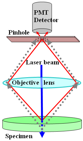
'Confocal' means having the same focal distance; pixels in a confocal image share the same focal plane in the studied specimen. Confocal microscopy permits observations of microscopic structures in thick specimens while normal microscopy cannot. Regular microscopy cannot resolve fine structures deep within a specimen,(which can be hundreds of microns thick,) because of light emitted and scattered by out-of-focus planes in the tissue. The formation of a confocal image is enabled by placing a barrier in the light path, with just a small pinhole in it allowing through only light rays originating in a single focal plane.

The laser beam used to generate fluorescence is focused by the microscope objective on a small spot in the specimen. Fluorescence emitted from this spot, still mixed with reflected light, is captured by the same objective lens. The reflected light is next filtered out by a dichroic mirror while the fluorescence proper continues on its way to the pinhole barrier.
The pinhole, or confocal aperture, is the next obstacle. Placed in front of the detector, it blocks all out-of-focus fluorescence emitted from either above or below the focal plane chosen, allowing only in-focus rays to pass through and be registered by the detector.
.A beam deflection mechanism now moves the laser from spot to spot, scanning the sample systematically in the selected x-y focal plane. Fluorescence emitted from each spot is measured in turn, generating a pixel-based, two dimensional image on the screen. This sharp image, which comes from a single focal plane is referred to as an optical section through the sample.
Additional optical sections can now be acquired by moving the stage up or down, and repeating the scanning process. A stepping motor adjusts the position of the focal plane on the Z axis in as little as 0.1 micron increments. A 3-D reconstruction of a specimen can be generated by stacking the 2-D optical sections collected in series.